Showing posts with label the brain. Show all posts

Monday, 6 September 2021

The Brain Doesn’t Think the Way You Think It Does

Brain Research 


 Familiar categories of mental functions such as perception, memory and attention reflect our experience of ourselves, but they are misleading about how the brain works. More revealing approaches are emerging.


Neuroscientists have tried to map various categories of mental function to specific regions of the brain, but recent work has shown that the definitions and boundaries of those regions are complex and context-dependent.

Lenka Šimečková for Quanta Magazine

Neuroscientists are the cartographers of the brain’s diverse domains and territories — the features and activities that define them, the roads and highways that connect them, and the boundaries that delineate them. Toward the front of the brain, just behind the forehead, is the prefrontal cortex, celebrated as the seat of judgment. Behind it lies the motor cortex, responsible for planning and coordinating movement. To the sides: the temporal lobes, crucial for memory and the processing of emotion. Above them, the somatosensory cortex; behind them, the visual cortex.

Not only do researchers often depict the brain and its functions much as mapmakers might draw nations on continents, but they do so “the way old-fashioned mapmakers” did, according to Lisa Feldman Barrett, a psychologist at Northeastern University. “They parse the brain in terms of what they’re interested in psychologically or mentally or behaviorally,” and then they assign the functions to different networks of neurons “as if they’re Lego blocks, as if there are firm boundaries there.”

But a brain map with neat borders is not just oversimplified — it’s misleading. “Scientists for over 100 years have searched fruitlessly for brain boundaries between thinking, feeling, deciding, remembering, moving and other everyday experiences,” Barrett said. A host of recent neurological studies further confirm that these mental categories “are poor guides for understanding how brains are structured or how they work.”

Neuroscientists generally agree about how the physical tissue of the brain is organized: into particular regions, networks, cell types. But when it comes to relating those to the task the brain might be performing — perception, memory, attention, emotion or action — “things get a lot more dodgy,” said David Poeppel, a neuroscientist at New York University.

No one disputes that the visual cortex enables sight, that the auditory cortex enables hearing, or that the hippocampus is essential for memory. Damage to those regions impairs those abilities, and researchers have identified mechanisms underlying them in those areas. But memory, for example, also requires brain networks other than the hippocampus, and the hippocampus is turning out to be key to a growing number of cognitive processes other than memory. Sometimes the degree of overlap is so great that the labels start to lose their meaning.

“The idea that there’s some kind of strong parallelism between mental categories that neuroscientists use to try and understand the brain and the neural implementation of mental events is just wrong,” Barrett said.

And while the current framework has led to important insights, “it’s gotten us stuck in certain traps that are really stifling research,” said Paul Cisek, a neuroscientist at the University of Montreal — an outcome that has also directly hobbled the development of treatments for neurological and psychological conditions.

That is why Barrett, Cisek and other scientists argue that for us to truly understand how the brain works, concepts at the field’s core may need to be revised, perhaps radically. As they grapple with that challenge, they are uncovering new ways to frame their questions about the brain, and new answers: This month alone, one such approach revealed an unexpected link between memory formation and metabolic regulation. But even if a new framework succeeds in explaining the brain’s operation, some researchers wonder whether the price of that success will be a loss of connection to our human experience.

‘More Aliases Than Sherlock Holmes’

When functional magnetic resonance imaging (fMRI) and other powerful technologies made it possible to examine living brains in increasingly sophisticated ways, neuroscientists enthusiastically started searching for the physical basis of our mental faculties. They made great strides in understanding the neural foundations of perception, attention, learning, memory, decision-making, motor control and other classic categories of mental activity.

But they also found unsettling evidence that those categories and the neural networks that support them don’t work as expected. It’s not just that the architecture of the brain disrespects the boundaries between the established mental categories. It’s that there’s so much overlap that a single brain network “has more aliases than Sherlock Holmes,” Barrett said.

Recent work has found, for instance, that two-thirds of the brain is involved in simple eye movements; meanwhile, half of the brain gets activated during respiration. In 2019, several teams of scientists found that most of the neural activity in “perception” areas such as the visual cortex was encoding information about the animals’ movements rather than sensory inputs.

This identity crisis isn’t limited to neural centers of perception or other cognitive functions. The cerebellum, a structure in the brains of all vertebrates, was thought to be dedicated almost exclusively to motor control, but scientists have found that it’s also instrumental in attention processes, the regulation of emotions, language processing and decision-making. The basal ganglia, another ancient part of the brain usually associated with motor control, has been similarly implicated in several high-level cognitive processes.

Some of these confusing results may come from methodological problems. To find where the human brain performs different functions, for instance, neuroscientists typically correlate cognitive processes with patterns of brain activity measured by fMRI. But studies suggest that researchers need to be more alert to irrelevant muscle twitches and fidgets that may contaminate the readings.

“You think that your results are telling you something about high-level cognition,” said György Buzsáki, a neuroscientist at the NYU School of Medicine, “when in fact, it may reflect nothing else except that, because of the task, [the subject’s] eyes are moving differently.”

But he and other scientists believe the recent findings also highlight a deeper conceptual problem in neuroscience. “We divide the real estate of the brain according to our preconceived ideas, assuming — wrongly, as far as I’m concerned — that those preconceived ideas have boundaries, and the same boundaries exist in brain function,” Buzsáki said.

In 2019, Russell Poldrack, a neuroscientist at Stanford University, and his colleagues set out to test how appropriate the recognized categories for mental function are. They gathered a massive amount of behavioral data — obtained from experiments designed to test different aspects of cognitive control, including working memory, response inhibition and learning — and ran it through a machine learning classifier. The resulting classifications defied expectations, mixing up traditional categories of brain results and sorting them into new groups that seemed to “move together in terms of some much more generic constructs,” Poldrack said — constructs for which we don’t yet have labels, and which might not relate directly to our conscious experience.

Another study by Poldrack’s colleagues found that tasks meant to measure either perception or memory “weren’t really measuring different constructs after all,” Poldrack said. “It suggests that those two categories are really imprecise.” It’s not that “perception” or “memory” is a useless term, he emphasized. But “if we want to understand what the brain does, we probably need much more precise ways to understand particular functions.”

The fact that it’s not even clear how to differentiate tests of perception from those of memory suggests that those categorical constructs “may not actually be the real organizing features of the mind,” Poldrack said.

Some scientists push back, arguing that so long as we know that the visual cortex isn’t just involved in vision, or that a memory network is doing more than its name suggests, we don’t necessarily need to rethink the categories themselves. But “sometimes an overly broad, vague use of a term can have detrimental effects on the types of experiments and hypotheses we generate,” said John Krakauer, a neuroscientist at Johns Hopkins University.

That’s perhaps been most obvious in research on emotions and mood.

Fear and Confusion

Joseph LeDoux is a neuroscientist at NYU known for his pioneering work on the amygdala, which is often referred to as the fear center of the brain. But that framing, he says, is very wrong — and very harmful. “I kept being introduced over the years as someone who discovered how feelings of fear come out of the amygdala,” he said. “But I would always kind of flinch when I would be introduced this way. Finally, I had enough.”

LeDoux has spent the past decade emphasizing that the amygdala isn’t involved in generating fear at all. Fear, he points out, is a cognitive interpretation of a situation, a subjective experience tied up in memory and other processes. The psychological phenomena that some people experience as fear may be experienced as something very different by others. Research shows that the feeling of fear arises in the prefrontal cortex and related brain areas.

The amygdala, on the other hand, is involved with processing and responding to threats — an ancient, subconscious behavioral and physiological mechanism. “The evidence shows that it’s not always fear that causes the behavior,” LeDoux said.

Calling the amygdala the fear center might seem innocuous, he continued, but “then the amygdala inherits all the semantic baggage of fear.” That mistake can distort attempts to develop medications, including those aiming to reduce anxiety. When potential treatments are tested in animals under stress, if the animals behave less timidly or show less physiological arousal, it’s usually interpreted as a reduction in anxiety or fear levels. But a medication can change someone’s behavioral or physiological responses — those outputs of the amygdala — without curing feelings of anxiety, LeDoux said.

“The whole field is suffering because of this confusion,” he said.

Similar problems occur in other areas, he added, such as studies of perception, where the physical processing of the sensory stimulus and the conscious experience of it are often bundled together. In both cases, LeDoux believes “these need to be pulled apart.”

Functional in Context

But teasing apart the significance of different brain areas is further complicated by the discovery that the involvement of neural systems in particular functions isn’t simply all or nothing. Sometimes it’s contingent on the details of what’s being processed.

Take the part of the medial temporal lobe called the perirhinal cortex — a crucial component of the classic “memory” system in the cortex. Elisabeth Murray of the National Institute of Mental Health and others did experiments in which humans and monkeys were asked to select a desired image from a pair that were morphed to resemble each other to varying degrees.

They found that the perirhinal cortex was involved in the performance of the task only when a particular amount of feature overlap was present. If the images were more similar or less, the perirhinal cortex had nothing to do with how well the humans or monkeys did. Similarly, the inferior temporal cortex, traditionally assigned a role in visual perception, was found to be crucial for memory tasks, but only in certain contexts.

To the retired neurobiologist Steven Wise, formerly of NIMH, the findings imply that instead of categorizing cortical areas in terms of their specialized visual, auditory, somatosensory or executive functions, researchers should study the different combinations of information they represent. One region might be involved in representing simple combinations of features, such as “orange” and “square” for an orange square. Other regions might have evolved to represent more complex combinations of visual features, or combinations of acoustic or quantitative information.

Wise argues that this brain organization scheme explains why there’s so much unexpected functional overlap in the traditional maps of mental activity. When each region represents a particular combination of information, “it does that for memory, and for perception, and for attention, and for the control of action,” Wise said.

That’s also why the perception and memory tasks that Murray used in her experiments only sometimes involved the perirhinal cortex: As the images in each task morphed, the combinations of features that distinguished them changed.

Wise’s representational framework is just one way of rethinking the brain’s subdivisions. While other researchers agree that the parts list guiding most neuroscientific research has problems, there’s little consensus about how to address it.

And even scientists in favor of a more radical rethinking of the field find it difficult to outline. “It’s easy to show how things are not working. The hard part now is where to go from here,” said Luiz Pessoa, a neuroscientist at the University of Maryland. “I’ve [often] caught myself using a whole lot of terms that I was criticizing the very use of. How can I say everything without saying ‘attention,’ ‘emotion,’ ‘motivation’?”

Cisek, in Montreal, is one of several researchers starting to rebuild the conceptual categories from an evolutionary perspective. For the past five years, he has been painstakingly making his way through vertebrate evolution, examining the progressive specialization of behavioral systems.

“Functional subdivisions do exist in the brain,” he said. “And they actually have an evolutionary history to them. If we could identify that history, it’ll help us identify the concepts better.”

Cisek has already used his new breakdown of brain activities to explain why, for instance, the basal ganglia plays a key role in some decision-making tasks but not others. “You realize that neither the term ‘decision-making’ nor the term ‘attention’ actually corresponds to a thing in the brain,” he said. “Instead, there are certain very pragmatic circuits in the brain, and they do certain things like ‘approach’ or ‘avoid.’ … Some of those things are going to look a bit like attention.”

Buzsáki takes a similar view. “We have to look at brain mechanisms first, and why and how those things evolved,” he said. For instance, memories, future planning and imagination are all partly encoded by the same neural mechanisms, which makes sense from an evolutionary perspective because the same system can be recycled for different purposes. “You may be better off thinking about all of [those] as one,” he said.

This approach is already leading to some intriguing discoveries. For years, Buzsáki has studied sharp wave-ripples, a type of brain activity in the hippocampus that enables the storage and retrieval of memories. But this month in Nature, his former doctoral student David Tingley and others in Buzsáki’s lab revealed an entirely new function for them: helping to regulate blood sugar levels.

“We are linking two very different extremes,” Buzsáki said — a basic metabolic process and a high-level cognitive one. He’s now hoping to uncover a deeper connection between the two, and to obtain insights into how sharp wave-ripples for body regulation might have been repurposed for memory formation.

Don’t Panic

Alternative approaches to studying mental categories are possible, too. Barrett, Pessoa and others, for instance, are considering whole-brain neural activity and an assortment of behaviors at the same time. “You study the whole system as its parts interact,” Barrett said. Functional categories such as memory, perception and attention can then be understood as “features of the brain state.”

Because of the counterintuitive groupings that emerged in his earlier study of behavioral data, Poldrack continues to be interested in model-free, data-driven searches for new categories. He thinks mental concepts could potentially be rewritten in computational terms — perhaps as a simplified version of the mathematical descriptions that define layers in artificial neural networks.

Each of these potential solutions has shortcomings. “But you don’t evaluate a new approach by all the questions it answers that the old one couldn’t,” Barrett said. “You evaluate it on the basis of what new questions does it stimulate.”

“There is no right way to do this,” she added. “There are only better ways and worse ways.”

Poldrack agreed. “I don’t think any of us would want to tell people: Don’t use the word ‘memory’ anymore,” he said. But to understand the brain, we might need to challenge our intuitions about how it works — “in the same way that quantum mechanics is challenging to comport with our understanding of physical phenomena in the world.”

Another important consideration is how meaningful a new framework might end up being. “You may gain in terms of knowledge, but you may actually stop understanding yourself,” Krakauer said.

When we wonder how the brain works, he explained, we want it to mean: What’s happening in my brain when I fall in love? Or when I’m excited? If we move too far away from our subjective experience and familiar cognitive concepts, he worries that what we learn about the brain might be like “42” in The Hitchhiker’s Guide to the Galaxy: the correct answer, but not to the question we had in mind. “Now, are we willing to live with that?” Krakauer asked.

Recent newsletters


Sunday, 29 August 2021

We have more than five senses. A neuroscientist explains the hidden abilities we often overlook

More than Five Senses 


  Photo by Solstice Hannan on Unsplash

Neuroscientist Dr Lisa Feldman Barrett delves into the different ways we’re able to perceive the world that go beyond sight, sound, touch, taste and smell.  


How many senses does the average human have? Assuming you equate senses with their receptors, such as the retinas in your eyes and the cochlea in your ears, then the traditional answer to this question is five – seeing, hearing, touch, smell and taste.

They’re called the ‘exteroceptive’ senses because they carry information about the external world.

But your body also has receptors for events occurring inside you, such as your beating heart, expanding lungs, gurgling stomach and many other movements that you’re completely unaware of. They’re traditionally grouped together as another sense, called ‘interoception’.


Yet a proper answer to this question is even more complex and interesting. For one thing, your body has receptors to carry other types of information, such as temperature, that we don’t usually consider to be senses.

Also, some of your receptors are used for more than one sense. Your retinas, for example, are portals for the light waves you need for vision, but some retinal cells also inform your brain if it’s daytime or nighttime. This unnamed ‘day/night sense’ is the basis for circadian rhythms that affect your metabolism and your sleep/wake cycle.


Read more about the science of senses:

Even senses that seem fundamental, such as vision, are intimately entwined with other senses that seem separate.

For example, it turns out that what you see, and how you see it, is yoked to your brain’s tracking of your heartbeat, which is part of interoception.

In the moments when your heart contracts and pushes blood out to your arteries, your brain takes in less visual information from the world.


Your brain also constructs senses that you don’t have receptors for. Examples are flavour, which the brain constructs from gustatory (taste) and olfactory (smell) data, and wetness, which is created from touch and temperature. 


In fact, your brain constructs everything you see, hear, smell, taste and feel using more than just the sense data from your body’s receptors. Light waves, for example, don’t simply enter your eyes, travel to your brain as electrical signals, and then you see.

Your brain actually predicts what you might see before you see it, based on past experience, the state of your body and your current situation. It combines its predictions with the incoming sense data from your retinas to construct your visual experience of the world around you.

Similarly, when you place your fingers on your wrist to feel your pulse, you’re actually feeling a construction based on your brain’s predictions and the actual sense data. You don’t experience sensations with your sense organs. You experience them with your brain.


Read more about neuroscience:


Tuesday, 20 July 2021

How Micro-Circuits in the Brain Regulate Fear

How Micro-Circuits in the Brain Regulate Fear


Photo by Frank Alarcon on Unsplash


16-Jul-2021 1:50 PM EDT, by University of Bern

Newswise — Fear is an important reaction that warns and protects us from danger. But when fear responses are out of control, this can lead to persistent fears and anxiety disorders. In Europe, about 15 percent of the population is affected by anxiety disorders. Existing therapies remain largely unspecific or are not generally effective, because the detailed neurobiological understanding of these disorders is lacking.

What was known so far is that distinct nerve cells interact together to regulate fear responses by promoting or suppressing them. Different circuits of nerve cells are involved in this process. A kind of "tug-of-war" takes place, with one brain circuit "winning" and overriding the other, depending on the context. If this system is disturbed, for example if fear reactions are no longer suppressed, this can lead to anxiety disorders.

Recent studies have shown that certain groups of neurons in the amygdala are crucial for the regulation of fear responses. The amygdala is a small almond-shaped brain structure in the center of the brain that receives information about fearful stimuli and transmits it to other brain regions to generate fear responses. This causes the body to release stress hormones, change heart rate or trigger fight, flight or freezing responses.

Now, a group led by Professors Stephane Ciocchi of the University of Bern and Andreas Luthi of the Friedrich Miescher Institute in Basel has discovered that the amygdala plays a much more active role in these processes than previously thought: Not only is the central amygdala a "hub" to generate fear responses, but it contains neuronal microcircuits that regulate the suppression of fear responses. In animal models, it has been shown that inhibition of these microcircuits leads to long-lasting fear behaviour. However, when they are activated, behaviour returns to normal despite previous fear responses. This shows that neurons in the central amygdala are highly adaptive and essential for suppressing fear. These results were published in the journal Nature Communications.

"Disturbed" suppression leads to long-lasting fear

The researchers led by Stephane Ciocchi and Andreas Luthi studied the activity of neurons of the central amygdala in mice during the suppression of fear responses. They were able to identify different cell types that influence the animals' behaviour. For their study, the researchers used several methods, including a technique called optogenetics with which they could precisely shut down - with pulses of light - the activity of an identified neuronal population within the central amygdala that produces a specific enzyme. This impaired the suppression of fear responses, whereupon animals became excessively fearful. "We were surprised how strongly our targeted intervention in specific cell types of the central amygdala affected fear responses," says Ciocchi, Assistant Professor at the Institute of Physiology, University of Bern. "The optogenetic silencing of these specific neurons completely abolished the suppression of fear and provoked a state of pathological fear."

Important for developing more effective therapies

In humans, dysfunction of this system, including deficient plasticity in the nerve cells of the central amygdala described here, could contribute to the impaired suppression of fear memories reported in patients with anxiety and trauma-related disorders. A better understanding of these processes will help develop more specific therapies for these disorders. "However, further studies are necessary to investigate whether discoveries obtained in simple animal models can be extrapolated to human anxiety disorders", Ciocchi adds.

This study was carried out in partnership with the University of Bern, the Friedrich Miescher Institute and international collaborators. It was funded by the University of Bern, the Swiss National Science Foundation and the European Research Council (ERC).

Systems Neuroscience Group, Institute of Physiology, University of Bern

Neuronal diversity is a hallmark of cortical networks. In the hippocampus, distinct neuronal cell-types interact together by selective synaptic contacts and neural activity patterns. We investigate how different forms of emotional and cognitive behaviours emerge within intricate neuronal circuits of the ventral CA1 hippocampus, a brain region instrumental for context-specific emotional memories, anxiety and goal-directed actions. We hypothesize that distinct behavioural programs are implemented by the selective recruitment of micro- and large-scale neural circuits of the ventral CA1 hippocampus. To identify these circuit motifs, we are combining single-unit recordings of ventral CA1 GABAergic interneurons and projection neurons, selective optogenetic strategies, cell-type specific viral tracing and behavioural paradigms in rodents. The results of our experimental approaches will determine fundamental neural computations underlying learning and memory within higher cortical brain regions.

###


Sunday, 4 July 2021

IQ tests can’t measure it, but ‘cognitive flexibility’ is key to learning and creativity

IQ Tests 


Photo by Riccardo Annandale on Unsplash


IQ is often hailed as a crucial driver of success, particularly in fields such as science, innovation and technology. In fact, many people have an endless fascination with the IQ scores of famous people. But the truth is that some of the greatest achievements by our species have primarily relied on qualities such as creativity, imagination, curiosity and empathy.

Many of these traits are embedded in what scientists call “cognitive flexibility” – a skill that enables us to switch between different concepts, or to adapt behaviour to achieve goals in a novel or changing environment. It is essentially about learning to learn and being able to be flexible about the way you learn. This includes changing strategies for optimal decision-making. In our ongoing research, we are trying to work out how people can best boost their cognitive flexibility.

Cognitive flexibility provides us with the ability to see that what we are doing is not leading to success and to make the appropriate changes to achieve it. If you normally take the same route to work, but there are now roadworks on your usual route, what do you do? Some people remain rigid and stick to the original plan, despite the delay. More flexible people adapt to the unexpected event and problem-solve to find a solution.

Cognitive flexibility may have affected how people coped with the pandemic lockdowns, which produced new challenges around work and schooling. Some of us found it easier than others to adapt our routines to do many activities from home. Such flexible people may also have changed these routines from time to time, trying to find better and more varied ways of going about their day. Others, however, struggled and ultimately became more rigid in their thinking. They stuck to the same routine activities, with little flexibility or change.

We want to ensure the knowledge of scientists reaches millions. Join us.

Huge advantages

Flexible thinking is key to creativity – in other words, the ability to think of new ideas, make novel connections between ideas, and make new inventions. It also supports academic and work skills such as problem solving. That said, unlike working memory – how much you can remember at a certain time – it is largely independent of IQ, or “crystallised intelligence”. For example, many visual artists may be of average intelligence, but highly creative and have produced masterpieces.

Contrary to many people’s beliefs, creativity is also important in science and innovation. For example, we have discovered that entrepreneurs who have created multiple companies are more cognitively flexible than managers of a similar age and IQ.

So does cognitive flexibility make people smarter in a way that isn’t always captured on IQ tests? We know that it leads to better “cold cognition”, which is non-emotional or “rational” thinking, throughout the lifespan. For example, for children it leads to better reading abilities and better school performance.

It can also help protect against a number of biases, such as confirmation bias. That’s because people who are cognitively flexible are better at recognising potential faults in themselves and using strategies to overcome these faults.

Cognitive flexibility is also associated with higher resilience to negative life events, as well as better quality of life in older individuals. It can even be beneficial in emotional and social cognition: studies have shown that cognitive flexibility has a strong link to the ability to understand the emotions, thoughts and intentions of others.

The opposite of cognitive flexibility is cognitive rigidity, which is found in a number of mental health disorders including obsessive-compulsive disorder, major depressive disorder and autism spectrum disorder.

Neuroimaging studies have shown that cognitive flexibility is dependent on a network of frontal and “striatal” brain regions. The frontal regions are associated with higher cognitive processes such as decision-making and problem solving. The striatal regions are instead linked with reward and motivation.

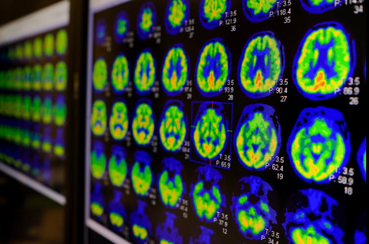
Image of brain scans.
Some people have more flexible brains. Utthapon wiratepsupon/Shutterstock

There are a number of ways to objectively assess people’s cognitive flexibility, including the Wisconsin Card Sorting Test and the CANTAB Intra-Extra Dimensional Set Shift Task.

Boosting flexibility

The good news is that it seems you can train cognitive flexibility. Cognitive behavioural therapy (CBT), for example, is an evidence-based psychological therapy which helps people change their patterns of thoughts and behaviour. For example, a person with depression who has not been contacted by a friend in a week may attribute this to the friend no longer liking them. In CBT, the goal is to reconstruct their thinking to consider more flexible options, such as the friend being busy or unable to contact them.

Structure learning – the ability to extract information about the structure of a complex environment and decipher initially incomprehensible streams of sensory information – is another potential way forward. We know that this type of learning involves similar frontal and striatal brain regions as cognitive flexibility.

In a collaboration between the University of Cambridge and Nanyang Technological University, we are currently working on a “real world” experiment to determine whether structural learning can actually lead to improved cognitive flexibility.

Studies have shown the benefits of training cognitive flexibility, for example in children with autism. After training cognitive flexibility, the children showed not only improved performance on cognitive tasks, but also improved social interaction and communication. In addition, cognitive flexibility training has been shown to be beneficial for children without autism and in older adults.

As we come out of the pandemic, we will need to ensure that in teaching and training new skills, people also learn to be cognitively flexible in their thinking. This will provide them with greater resilience and wellbeing in the future.

Cognitive flexibility is essential for society to flourish. It can help maximise the potential of individuals to create innovative ideas and creative inventions. Ultimately, it is such qualities we need to solve the big challenges of today, including global warming, preservation of the natural world, clean and sustainable energy and food security.

Professors Trevor Robbins, Annabel Chen and Zoe Kourtzi also contributed to this article.


Friday, 4 June 2021

What Happens to Our Brains When We Get Depressed?

Photo by The National Cancer Institute on Unsplash

The Brain 


The human brain, in all its complexity, is nearly impossible to model. One neuroscientist is trying anyway


Wednesday, 9 December 2020

Science as We Know It Can’t Explain Consciousness – but a Revolution Is Coming

Our Minds 


One day there will be a science of consciousness, but it won’t look like science does now.


Explaining how something as complex as consciousness can emerge from a grey, jelly-like lump of tissue in the head is arguably the greatest scientific challenge of our time. The brain is an extraordinarily complex organ, consisting of almost 100 billion cells – known as neurons – each connected to 10,000 others, yielding some ten trillion nerve connections.

We have made a great deal of progress in understanding brain activity, and how it contributes to human behaviour. But what no one has so far managed to explain is how all of this results in feelings, emotions and experiences. How does the passing around of electrical and chemical signals between neurons result in a feeling of pain or an experience of red?

There is growing suspicion that conventional scientific methods will never be able answer these questions. Luckily, there is an alternative approach that may ultimately be able to crack the mystery.

For much of the 20th century, there was a great taboo against querying the mysterious inner world of consciousness – it was not taken to be a fitting topic for “serious science”. Things have changed a lot, and there is now broad agreement that the problem of consciousness is a serious scientific issue. But many consciousness researchers underestimate the depth of the challenge, believing that we just need to continue examining the physical structures of the brain to work out how they produce consciousness.

The problem of consciousness, however, is radically unlike any other scientific problem. One reason is that consciousness is unobservable. You can’t look inside someone’s head and see their feelings and experiences. If we were just going off what we can observe from a third-person perspective, we would have no grounds for postulating consciousness at all.

Of course, scientists are used to dealing with unobservables. Electrons, for example, are too small to be seen. But scientists postulate unobservable entities in order to explain what we observe, such as lightning or vapour trails in cloud chambers. But in the unique case of consciousness, the thing to be explained cannot be observed. We know that consciousness exists not through experiments but through our immediate awareness of our feelings and experiences.

file-20191031-187925-xzj4re.jpg

Only you can experience your emotions. Photo by Olga Danylenko.

So how can science ever explain it? When we are dealing with the data of observation, we can do experiments to test whether what we observe matches what the theory predicts. But when we are dealing with the unobservable data of consciousness, this methodology breaks down. The best scientists are able to do is to correlate unobservable experiences with observable processes, by and relying on their reports regarding their private conscious experiences.

By this method, we can establish, for example, that the invisible feeling of hunger is correlated with visible activity in the brain’s hypothalamus. But the accumulation of such correlations does not amount to a theory of consciousness. What we ultimately want is to explain why conscious experiences are correlated with brain activity. Why is it that such activity in the hypothalamus comes along with a feeling of hunger?

In fact, we should not be surprised that our standard scientific method struggles to deal with consciousness. As I explore in my new book, Galileo’s Error: Foundations for a New Science of Consciousness, modern science was explicitly designed to exclude consciousness.

Before the “father of modern science” Galileo Galilei, scientists believed that the physical world was filled with qualities, such as colours and smells. But Galileo wanted a purely quantitative science of the physical world, and he therefore proposed that these qualities were not really in the physical world but in consciousness, which he stipulated was outside of the domain of science.

This worldview forms the backdrop of science to this day. And so long as we work within it, the best we can do is to establish correlations between the quantitative brain processes we can see and the qualitative experiences that we can’t, with no way of explaining why they go together.

Mind is Matter

I believe there is a way forward, an approach that’s rooted in work from the 1920s by the philosopher Bertrand Russell and the scientist Arthur Eddington. Their starting point was that physical science doesn’t really tell us what matter is.

This may seem bizarre, but it turns out that physics is confined to telling us about the behaviour of matter. For example, matter has mass and charge, properties which are entirely characterised in terms of behaviour – attraction, repulsion and resistance to acceleration. Physics tells us nothing about what philosophers like to call “the intrinsic nature of matter”, how matter is in and of itself.

It turns out, then, that there is a huge hole in our scientific world view – physics leaves us completely in the dark about what matter really is. The proposal of Russell and Eddington was to fill that hole with consciousness.

The result is a type of “panpsychism” – an ancient view that consciousness is a fundamental and ubiquitous feature of the physical world. But the “new wave” of panpsychism lacks the mystical connotations of previous forms of the view. There is only matter – nothing spiritual or supernatural – but matter can be described from two perspectives. Physical science describes matter “from the outside”, in terms of its behaviour, but matter “from the inside” is constituted of forms of consciousness.

This means that mind is matter, and that even elementary particles exhibit incredibly basic forms of consciousness. Before you write that off, consider this. Consciousness can vary in complexity. We have good reason to think that the conscious experiences of a horse are much less complex than those of a human being, and that the conscious experiences of a rabbit are less sophisticated than those of a horse. As organisms become simpler, there may be a point where consciousness suddenly switches off – but it’s also possible that it just fades but never disappears completely, meaning even an electron has a tiny element of consciousness.

What panpsychism offers us is a simple, elegant way of integrating consciousness into our scientific worldview. Strictly speaking it cannot be tested; the unobservable nature of consciousness entails that any theory of consciousness that goes beyond mere correlations is not strictly speaking testable. But I believe it can be justified by a form of inference to the best explanation: panpsychism is the simplest theory of how consciousness fits in to our scientific story.

While our current scientific approach offers no theory at all – only correlations – the traditional alternative of claiming that consciousness is in the soul leads to a profligate picture of nature in which mind and body are distinct. Panpsychism avoids both of these extremes, and this is why some of our leading neuroscientists are now embracing it as the best framework for building a science of consciousness.

I am optimistic that we will one day have a science of consciousness, but it won’t be science as we know it today. Nothing less than a revolution is called for, and it’s already on its way.

Philip Goff is an Assistant Professor of Philosophy at Durham University.

The Conversation

Sunday, 29 November 2020

Exercise and the brain: three ways physical activity changes its very structure

The Outdoors 

 Having been in indoors for two weeks because someone in my family tested positive to Corona Virus, I am longing to get out and walk and sample the fresh air once more.


Give a shoutout to Lital Levy on social or copy the text below to attribute.

Regular exercise changes the structure of our bodies’ tissues in obvious ways, such as reducing the size of fat stores and increasing muscle mass. Less visible, but perhaps even more important, is the profound influence exercise has on the structure of our brains – an influence that can protect and preserve brain health and function throughout life. In fact, some experts believe that the human brain may depend on regular physical activity to function optimally throughout our lifetime.

Here are just a few ways exercise changes the structure of our brain.

Memory

Many studies suggest that exercise can help protect our memory as we age. This is because exercise has been shown to prevent the loss of total brain volume (which can lead to lower cognitive function), as well as preventing shrinkage in specific brain regions associated with memory. For example, one magnetic resonance imaging (MRI) scan study revealed that in older adults, six months of exercise training increases brain volume.

Another study showed that shrinkage of the hippocampus (a brain region essential for learning and memory) in older people can be reversed by regular walking. This change was accompanied by improved memory function and an increase of the protein brain-derived neutropic factor (BDNF) in the bloodstream.

Fight back against disinformation. Get your news here, direct from experts

BDNF is essential for healthy cognitive function due to its roles in cell survival, plasticity (the brain’s ability to change and adapt from experience) and function. Positive links between exercise, BDNF and memory have been widely investigated and have been demonstrated in young adults and older people.

BDNF is also one of several proteins linked with adult neurogenesis, the brain’s ability to modify its structure by developing new neurons throughout adulthood. Neurogenesis occurs only in very few brain regions – one of which is the hippocampus – and thus may be a central mechanism involved in learning and memory. Regular physical activity may protect memory in the long term by inducing neurogenesis via BDNF.

While this link between exercise, BDNF, neurogenesis, and memory is very well described in animal models, experimental and ethical constraints mean that its importance to human brain function is not quite so clear. Nevertheless exercise-induced neurogenesis is being actively researched as a potential therapy for neurological and psychiatric disorders, such as Alzheimer’s disease, Parkinson’s disease and depression.

Blood vessels

The brain is highly dependent on blood flow, receiving approximately 15% of the body’s entire supply – despite being only 2-3% of our body’s total mass. This is because our nervous tissues need a constant supply of oxygen to function and survive. When neurons become more active, blood flow in the region where these neurons are located increases to meet demand. As such, maintaining a healthy brain depends on maintaining a healthy network of blood vessels.

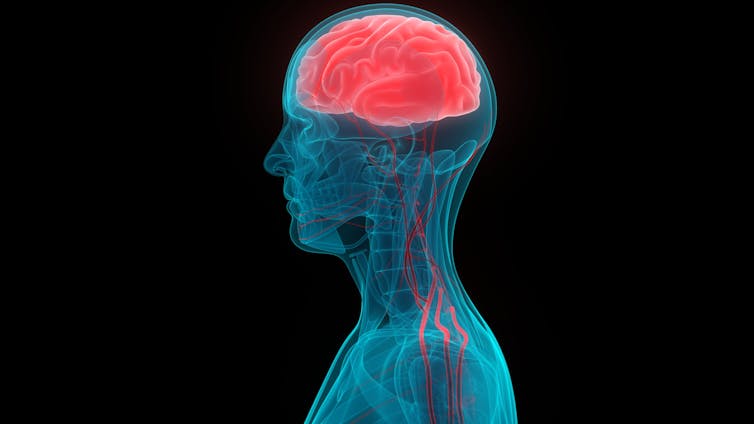
An illustration of a person's brain and the blood vessels connected to it.
Regular exercise helps blood vessels grow in the brain. Magic mine/ Shutterstock

Regular exercise increases the growth of new blood vessels in the brain regions where neurogenesis occurs, providing the increased blood supply that supports the development of these new neurons. Exercise also improves the health and function of existing blood vessels, ensuring that brain tissue consistently receives adequate blood supply to meet its needs and preserve its function.

Finally, regular exercise can prevent, and even treat, hypertension (high blood pressure), which is a risk factor for development of dementia. Exercise works in multiple ways to enhance the health and function of blood vessels in the brain.

Inflammation

Recently, a growing body of research has centred on microglia, which are the resident immune cells of the brain. Their main function is to constantly check the brain for potential threats from microbes or dying or damaged cells, and to clear any damage they find.


Read more: Microglia: the brain’s 'immune cells' protect against diseases – but they can also cause them


With age, normal immune function declines and chronic, low-level inflammation occurs in body organs, including the brain, where it increases risk of neurodegenerative disease, such as Alzheimer’s disease. As we age, microglia become less efficient at clearing damage, and less able to prevent disease and inflammation. This means neuroinflammation can progress, impairing brain functions – including memory.

But recently, we’ve shown that exercise can reprogramme these microglia in the aged brain. Exercise was shown to make the microglia more energy efficient and capable of counteracting neuroinflammatory changes that impair brain function. Exercise can also modulate neuroinflammation in degenerative conditions like Alzheimer’s disease and multiple sclerosis. This shows us the effects of physical activity on immune function may be an important target for therapy and disease prevention.

So how can we ensure that we’re doing the right kind of exercise – or getting enough of it – to protect the brain? As yet, we don’t have robust enough evidence to develop specific guidelines for brain health though findings to date suggest that the greatest benefits are to be gained by aerobic exercise – such as walking, running, or cycling. It’s recommended adults get a minimum of 150 minutes per week of moderate intensity aerobic exercise, combined with activities that maintain strength and flexibility, to maintain good general health.

It must also be noted that researchers don’t always find exercise has beneficial effect on the brain in their studies – likely because different studies use different exercise training programmes and measures of cognitive function, making it difficult to directly compare studies and results. But regardless, plenty of research shows us that exercise is beneficial for many aspects of our health, so it’s important to make sure you’re getting enough. We need to be conscious of making time in our day to be active – our brains will thank us for it in years to come.